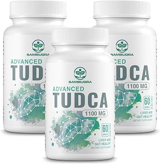
Tauroursodeoxycholic acid (TUDCA) is a naturally occurring, water-soluble bile acid formed when ursodeoxycholic acid (UDCA) is conjugated with taurine. Long studied in the context of cholestatic liver disease, TUDCA has drawn growing scientific interest for a more fundamental reason: its apparent ability to interfere with the intrinsic, or mitochondrial, pathway of programmed cell death. Understanding how a bile acid derivative can suppress apoptosis at the organelle level requires a brief look at why mitochondria are the central decision point in cell fate.
Inside stressed or damaged cells, the mitochondrion serves as a gatekeeper of death signals. When pro-apoptotic proteins breach the outer mitochondrial membrane, cytochrome c pours into the cytoplasm and triggers a caspase cascade that dismantles the cell from within. TUDCA appears to interrupt this sequence at several points — from stabilizing the outer membrane against pore-forming proteins, to modulating the survival signals that keep pro-death proteins in check. This article examines the proposed mechanisms in detail, drawing exclusively on the available peer-reviewed evidence.
Key Takeaways
- TUDCA blocks Bax from permeabilizing the outer mitochondrial membrane, preventing cytochrome c release before the caspase cascade can begin [1].
- It also suppresses downstream pro-apoptotic proteins including Bad and engages the PI3K survival-kinase pathway, indicating multi-node antiapoptotic activity [2] [3].
- Hydrophobic bile acids damage mitochondria through ROS generation and mPTP opening; TUDCA’s hydrophilicity may partly counteract this in relevant disease contexts [4] [5].
- Protective effects have been studied across hepatic, neuronal, and cardiac cell models, but large-scale human trial data on mitochondrial apoptosis outcomes outside of cholestasis are not available.
- Context and cell type matter: closely related bile acid derivatives can behave differently across cell types, and findings from one experimental model should not be generalized without further evidence [6].
The Intrinsic Apoptosis Pathway: Why Mitochondria Are the Decision Point
The intrinsic apoptosis pathway is activated by internal stressors — DNA damage, oxidative injury, misfolded proteins, or toxic metabolites. These stimuli shift the balance between pro-apoptotic proteins (principally Bax and Bak) and anti-apoptotic ones (Bcl-2, Bcl-xL). When pro-apoptotic proteins prevail, they oligomerize and form pores in the outer mitochondrial membrane, causing mitochondrial outer membrane permeabilization (MOMP). This allows cytochrome c and other apoptogenic factors to exit the intermembrane space and enter the cytosol.
Once released, cytochrome c binds Apaf-1 and pro-caspase-9 to form the apoptosome, which cleaves and activates executioner caspases — ultimately causing DNA fragmentation and cell death. Separately, the mitochondrial permeability transition pore (mPTP) can open under extreme stress, collapsing membrane potential and triggering a less orderly form of cell death. Research tracking the progressive stages of mitochondrial destruction caused by cell-toxic bile salts has helped illuminate both routes in detail [5], providing an important cellular model for studying what protective agents like TUDCA must counteract.
How Hydrophobic Bile Acids Destabilize Mitochondria
Not all bile acids are equally benign. Hydrophobic, detergent-like bile salts can directly attack mitochondrial membranes, uncouple oxidative phosphorylation, and generate reactive oxygen species (ROS). Research using human hepatic mitochondria demonstrated that exposure to hydrophobic bile acids causes both ROS generation and opening of the mitochondrial permeability transition pore [4]. This creates a vicious cycle: oxidative stress further destabilizes the membrane, producing more ROS and accelerating the collapse of membrane potential.
Cell biology studies have mapped progressive stages of this mitochondrial destruction, showing that cell-toxic bile salts cause sequential swelling, cristae remodeling, and membrane rupture [5]. The downstream consequences extend beyond acute cell death: disrupted mitochondrial enzyme activity has been linked to hepatic lipid accumulation, as illustrated by work on mitochondrial glycerol 3-phosphate dehydrogenase deficiency [7]. This body of work explains why preserving mitochondrial integrity is a credible therapeutic target, particularly in tissues with high metabolic demand such as the liver, heart, and nervous system. TUDCA, being highly hydrophilic, does not share the membrane-destabilizing properties of its toxic counterparts.

TUDCA's Core Mechanism: Stabilizing the Outer Membrane Against Bax
The most direct evidence that TUDCA intervenes in the mitochondrial death pathway comes from experiments using isolated mitochondria. TUDCA was shown to prevent Bax-induced membrane perturbation and cytochrome c release in isolated mitochondria, acting upstream of the full apoptotic cascade [1]. This is mechanistically significant: by limiting the membrane-permeabilizing activity of Bax, TUDCA may prevent the very step that commits a cell to apoptosis before caspases are ever activated.

Complementary work in human hepatoma (HepG2) cells examined cytochrome c-mediated apoptosis in a hepatic cell model, finding that TUDCA exerted protective effects against cytochrome c-triggered death signaling [8]. Together, these studies suggest TUDCA acts at more than one node in the apoptotic sequence — both at the outer membrane where cytochrome c escape begins, and further downstream where released cytochrome c drives caspase activation.
It is worth noting that not every bile acid derivative behaves the same way. Ursodeoxycholic acid (UDCA), the unconjugated parent compound, has been shown to induce apoptosis in human melanoma cells through the mitochondrial pathway [6] — an effect that may be desirable in cancer contexts but illustrates that closely related molecules can have cell-type-dependent and context-dependent effects. The taurine conjugation distinguishing TUDCA from UDCA alters its physicochemical properties and biological activity, and findings from one should not be uncritically applied to the other.
Downstream Antiapoptotic Signals: Bad Inhibition and the PI3K Pathway
TUDCA’s antiapoptotic reach extends beyond the outer mitochondrial membrane. In hepatocytes expressing the misfolding-prone Z variant of alpha-1 antitrypsin — a protein whose intracellular accumulation triggers progressive liver injury — TUDCA inhibited apoptosis through suppression of the pro-apoptotic protein Bad [2]. Bad, when unphosphorylated, sequesters Bcl-2 and Bcl-xL, freeing Bax to act. By keeping Bad in check, TUDCA preserves the anti-apoptotic buffer that restrains Bax-driven membrane permeabilization.
In neuronal cells threatened by amyloid-beta peptide, TUDCA prevented cell death via a phosphatidylinositol 3-kinase (PI3K)-dependent signaling pathway [3]. PI3K signaling activates Akt, which phosphorylates and inactivates Bad, reinforcing Bcl-2 survival signals. This finding suggests TUDCA may engage active survival kinase cascades rather than simply blocking death signals passively — an important mechanistic distinction.
Work using 3-nitropropionic acid, a mitochondrial toxin that inhibits complex II of the electron transport chain, further demonstrated that TUDCA can partially prevent apoptosis through a mitochondrial pathway that is independent of the permeability transition pore itself [9]. This mPTP-independent protection implies TUDCA’s intervention does not rely solely on preventing pore opening — it engages additional stabilizing mechanisms that are not yet fully characterized.
Broader Tissue Contexts: Heart, Liver, and the ER Stress Connection
While much of the foundational mechanistic work was done in liver and neuronal models, evidence suggests mitochondria-protective effects may extend to cardiac tissue. In a high-fat diet mouse model, TUDCA mitigated cardiomyocyte contractile dysfunction and abnormal intracellular calcium handling — changes associated with mitochondrial and ER stress in the context of metabolic overload [10]. Calcium dysregulation is a well-established trigger of mPTP opening, so normalizing Ca2+ dynamics may represent an indirect mechanism by which TUDCA shields the mitochondrial membrane.

The connection to endoplasmic reticulum (ER) stress is also relevant here. The ER and mitochondria are physically and functionally coupled through mitochondria-associated membranes. When the ER is overwhelmed by misfolded proteins, its stress signals propagate to the mitochondrion, promoting pro-apoptotic Bcl-2 family protein activation and cytochrome c release. Research has demonstrated that inhibiting ER stress protects both steatotic and non-steatotic livers from apoptosis during ischemia-reperfusion injury [11], providing a tissue-level rationale for compounds that target ER stress upstream of mitochondrial involvement. Other stressors that simultaneously drive ER stress and mitochondrial dysfunction can potently activate the apoptosis cascade through both organelles concurrently [12], underscoring how tightly interconnected these two death-signaling hubs are — and why a molecule capable of acting at both may offer broader protection.
What the Evidence Does and Does Not Establish
The mechanistic picture above is largely derived from in vitro cell culture experiments and animal models. Studies in isolated mitochondria [1] and hepatoma cell lines [8] are valuable for identifying biochemical mechanisms, but they do not automatically translate to outcomes in living humans taking oral TUDCA. Oral TUDCA undergoes hepatic first-pass metabolism and enterohepatic recirculation; the concentrations reaching target tissues outside the liver — neurons, cardiomyocytes — are not well characterized at the doses used in human supplementation contexts.
Clinically, TUDCA’s strongest evidence base remains in cholestatic liver disease, where it improves bile flow and liver enzyme profiles. Evidence for mitochondria-protective effects in healthy humans — preventing cellular apoptosis in the absence of an underlying disease state — is not established by large-scale randomized controlled trials. The existing studies represent important mechanistic groundwork, but the gap between isolated mitochondria and a healthy human liver or brain is substantial. Interpreting these findings with appropriate caution is not pessimism; it is the standard demanded by the evidence.
🛒 Where to Buy TUDCA
- BulkSupplements.com TUDCA CapsulesLab-tested / studied
capsules, 500 mg per capsule, 90 capsules — Third-party tested and made in a cGMP facility; one capsule per serving; higher per-capsule dose suits users targeting 500–1000 mg/day protocols - Nutricost TUDCA 250mg
capsules, 250 mg per capsule, 60 capsules — High-volume seller; non-GMO and gluten-free labeling; no third-party purity COA publicly posted, but consistent community reputation for accurate dosing - Double Wood Supplements TUDCA Bile Salts 500mg
capsules, 500 mg per capsule, 60 capsules — USA-manufactured; publishes basic COA on request; popular among biohacker community for reliable potency at accessible price point - Nutricost TUDCA Powder
powder, 25 g tub — Best cost-per-gram option for daily high-dose users; gluten-free, same material as their capsule line; requires a milligram-accurate scale for precise dosing
As an Amazon Associate we earn from qualifying purchases. TUDCA quality varies widely — always choose a product that publishes a third-party purity test (COA) confirming 98%+ tauroursodeoxycholic acid before buying.
A Note on the Evidence
The evidence reviewed here is predominantly from in vitro and animal studies; robust large-scale randomized controlled trials demonstrating mitochondria-protective outcomes in healthy humans are not yet available, and TUDCA is contraindicated in bile duct obstruction, should be used with caution in patients with gallbladder disease, cholangitis, or severe hepatic impairment, and may interact with bile acid sequestrants, cyclosporine, and certain lipid-lowering agents — consult a qualified healthcare provider before use.
Frequently Asked Questions
How does TUDCA prevent cytochrome c release from mitochondria?
The primary mechanism identified in laboratory research is TUDCA’s ability to prevent Bax from permeabilizing the outer mitochondrial membrane. In experiments using isolated mitochondria, TUDCA prevented Bax-induced membrane perturbation and the cytochrome c release that follows [1]. Without cytochrome c reaching the cytosol, the apoptosome cannot form and the downstream caspase cascade is not initiated.
Does TUDCA act through a single mechanism or multiple antiapoptotic pathways?
Research points to multiple mechanisms acting in parallel. Beyond blocking Bax at the outer membrane [1], TUDCA has been shown to suppress the pro-apoptotic protein Bad [2], activate PI3K-dependent survival signaling [3], and confer protection independent of the mitochondrial permeability transition pore under certain toxic stressors [9]. This multi-node activity may partly explain why protective effects appear across different cell stress models.
Why are hydrophobic bile acids particularly damaging to mitochondria?
Hydrophobic bile salts act in a detergent-like fashion that disrupts biological membranes. In human hepatic mitochondria, exposure to these bile acids causes reactive oxygen species generation and triggers opening of the mitochondrial permeability transition pore [4]. Studies tracking the progression of this damage show sequential mitochondrial swelling, structural disruption, and eventual membrane rupture [5]. TUDCA, being highly hydrophilic, does not share these membrane-destabilizing properties.
Can TUDCA protect neurons as well as liver cells?
Cell-based studies suggest neuroprotective activity in specific experimental contexts. TUDCA protected neuronal cells from amyloid-beta-induced death via a PI3K-dependent pathway [3] and partially prevented apoptosis triggered by a mitochondrial toxin through a mechanism independent of permeability transition pore opening [9]. These findings are mechanistically interesting but preliminary; clinical neuroprotective effects in humans have not been established by large randomized controlled trials.
Is there any cardiac relevance to TUDCA's mitochondrial activity?
Animal model research has found that TUDCA can mitigate cardiomyocyte contractile dysfunction and abnormal intracellular calcium handling caused by a high-fat diet [10]. Because calcium overload is a known trigger of mitochondrial permeability transition, normalizing Ca2+ dynamics may represent one indirect mechanism linking TUDCA to cardiac mitochondrial stability. This remains exploratory research and has not been tested in human cardiac clinical trials.
Is TUDCA always antiapoptotic, or does it depend on the cell type?
Context and cell type matter considerably. Most mechanistic research describes TUDCA as protecting normal cells from stress-induced apoptosis. However, closely related molecules can behave differently across cell types: UDCA, the unconjugated parent compound, has been shown to induce apoptosis in human melanoma cells through the mitochondrial pathway [6] — an entirely different therapeutic intent. TUDCA’s antiapoptotic profile in healthy cells should not be assumed to generalize universally without additional targeted research.
References
- Rodrigues CM et al. Tauroursodeoxycholic acid prevents Bax-induced membrane perturbation and cytochrome C release in isolated mitochondria. Biochemistry (2003). PMID 12627974
- Miller SD et al. Tauroursodeoxycholic acid inhibits apoptosis induced by Z alpha-1 antitrypsin via inhibition of Bad. Hepatology (Baltimore, Md.) (2007). PMID 17559149
- Solá S et al. Tauroursodeoxycholic acid prevents amyloid-beta peptide-induced neuronal death via a phosphatidylinositol 3-kinase-dependent signaling pathway. Molecular medicine (Cambridge, Mass.) (2003). PMID 15208744
- Sokol RJ et al. Human hepatic mitochondria generate reactive oxygen species and undergo the permeability transition in response to hydrophobic bile acids. Journal of pediatric gastroenterology and nutrition (2005). PMID 16056106
- Schulz S et al. Progressive stages of mitochondrial destruction caused by cell toxic bile salts. Biochimica et biophysica acta (2013). PMID 23685124
- Yu H et al. Apoptosis induced by ursodeoxycholic acid in human melanoma cells through the mitochondrial pathway. Oncology reports (2019). PMID 30542709
- Zheng Y et al. Deficiency of Mitochondrial Glycerol 3-Phosphate Dehydrogenase Contributes to Hepatic Steatosis. Hepatology (Baltimore, Md.) (2019). PMID 30653687
- Xie Q et al. [Effect of Tauroursodeoxycholic acid on cytochrome C-mediated apoptosis in HepG2 cells]. Zhonghua gan zang bing za zhi = Zhonghua ganzangbing zazhi = Chinese journal of hepatology (2003). PMID 12773247
- Rodrigues CM et al. Tauroursodeoxycholic acid partially prevents apoptosis induced by 3-nitropropionic acid: evidence for a mitochondrial pathway independent of the permeability transition. Journal of neurochemistry (2000). PMID 11080188
- Turdi S et al. Tauroursodeoxycholic acid mitigates high fat diet-induced cardiomyocyte contractile and intracellular Ca2+ anomalies. PloS one (2013). PMID 23667647
- Ben Mosbah I et al. Endoplasmic reticulum stress inhibition protects steatotic and non-steatotic livers in partial hepatectomy under ischemia-reperfusion. Cell death & disease (2010). PMID 21364657
- Zhou L et al. Miltirone exhibits antileukemic activity by ROS-mediated endoplasmic reticulum stress and mitochondrial dysfunction pathways. Scientific reports (2016). PMID 26848099
These statements have not been evaluated by the Food and Drug Administration. This information is not intended to diagnose, treat, cure, or prevent any disease. Content is for informational purposes only and is not medical advice; consult a qualified healthcare provider before starting any supplement. As an Amazon Associate we earn from qualifying purchases.